|

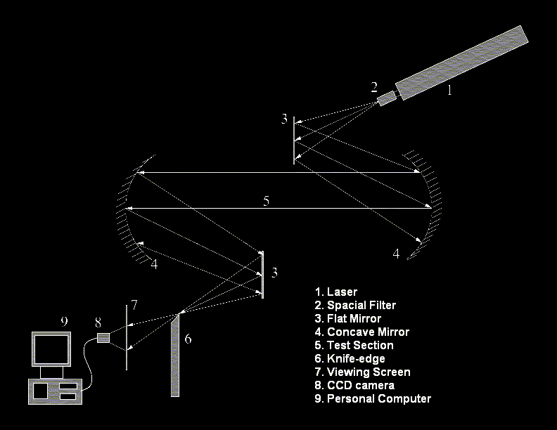
Figure at the right shows the schematic diagram of a Z-type schlieren set-up. Lase beam from the laser source (1) is
collimated by a mirror system (4). The collimated beam passes via the test section (5) and focussed back using another mirror
system (4). A knife-edge (6) is placed at the focus of the beam. The image is formed at the screen (7) which is captured by
an CCD camera (8) and the image is transferred to the computer (9) for further processing.

The schlieren technique works on the principle of deflection of light beam. The knife edge plays a crutial role in defining
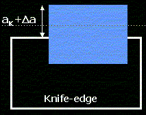
the contrast of the image. In fact no information can be extracted without the presence of knife edge. Figure below left shows
the initial knife edge setting. The knife edge blocks most of the light beam to give a very dark initial image. Slight amount
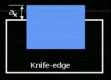
of light (ak) is allowed to pass to give an initial image. Figure below right shows the final position of light beam with
respect to the knife edge. Due to variation of refractive index with the presence of thermal disturbance, the light beam gets
deflected upwards (or may be downwards) and more (or less) light passes over the knife edge (ak + delta a). This results
in change in illumination st the screen giving a different image.


The difference in the contrast of the image gives the first differencial of the refractive index gradient. Integration
of the contrast with known boundary condition can provide quantitaive information on the thermal field.
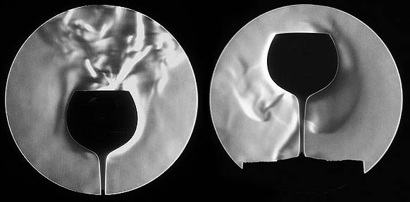
Image below shows a typical schlieren image for hot and cold cup.

The hot cup (left) reduces the density of adjacent air whcih becomes lighter and moves up. The cold cup (right) makes
the air dense and heavy which moves down.
Schlieren is a widely used qualitative visualization technique. But it cal also be applied for quantitative data extraction
for simple flows with well known boundary conditions. It can then also be advanced to get 3-d data using tomographic techniques.
|
